This is an online e log book to
discuss our patient de-identified health data shared after taking his /
her / guardians signed informed consent. Here we discuss our individual
patients problems through series of inputs from available global online
community of experts with an aim to solve those patients clinical
problem with collective current best evident based input.
This E blog also reflects my patient centered online learning portfolio and your valuable inputs on the comment box is welcome.
I
have been given this case to solve in an attempt to understand the
topic of " patient clinical data analysis" to develop my competency in
reading and comprehending clinical data including history, clinical
findings, investigations and come up with diagnosis and treatment plan.
A 62 year old male came to the OPD with chief complaints of
CHIEF COMPLAINTS:
Pedal edema since 1 month
Decreased urine output since 1 month
Fever since 3 days
HISTORY OF PRESENTING ILLNESS:
Patient
was apparently asymptomatic 3 years back then developed pedal edema,
shortness of breath, fever, cough and was admitted in a private hospital
hyd and diagnosed as renal failure.
In
February 2022 patient came to kamineni Narketpalli with chief complaints
of shortness of breath and decreased appetite and undergone dialysis
under 3 sessions and was on conservative management.
In November 2022, patient came with similar complaints and undergone dialysis here
Now
he developed pedal edema since 1 month which is pitting type and
complained of fever since 3 days which is continuous ,high grade and
associated with chills and rigor.
H/o nausea, vomiting, anorexia on 4th jan night.
Vomiting is non projectile, non bilious, non blood tinged contained food particles associated with nausea in 2-3 episodes.
H/o decreased urine output since 1month
No h/o burning micturition, pain abdomen.
H/o abscess over left medial and infra gluteal region 1 year back.
Came for dialysis ( no regular follow up)
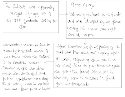
Timeline of events:
PAST HISTORY:
K/C/O Diabetes since 3 years
K/C/O Hypertension since 3 years and on medication for both
Patient had a history of knee injury 3 years back for which he undergone surgery.
N/K/C/O CAD, epilepsy, asthma, Tuberculosis.
No history of any blood transfusions.
FAMILY HISTORY: No significant family history
PERSONAL HISTORY:
DIET: Mixed
APPETITE: Decreased
SLEEP: Adequate
BOWEL MOVEMENTS: Regular
BLADDER MOVEMENTS: Decreased urine output
ADDICTIONS: Drinks toddy occasionally
GENERAL EXAMINATION:
Patient is conscious, coherent and cooperative Well oriented to time, place and person
Moderately built and moderately nourished.
Pallor-absent
Icterus-absent
Cyanosis-absent
Clubbing-absent
Lymphadenopathy-absent
Pedal edema-present
VITALS:. .
Temp:Febrile(102°F)
Blood pressure:130/90mmHg
Pulse rate:82bpm
Respiratory rate:14cpm
SYSTEMIC EXAMINATION.
CVS EXAMINATION :-
JVP: Normal
INSPECTION:
Chest wall symmetrical
Pulsations not seen
PALPATION:
Apical impulse – normal
Pulsations – normal
Thrills absent
PERCUSSION:
No abnormal findings
AUSCULTATION:
S1, S2 heard
No murmurs
No added sounds
RESPIRATORY EXAMINATION :-
- Chest bilaterally symmetrical, all quadrants
moves equally with respiration.
- Trachea central, chest expansion normal.
- Resonant on percussion
- Bilateral equal air entry, no added sounds heard.
1. Breath sounds - Normal Vesicular Breath sounds
2. Added sounds - absent
3. Vocal Resonance - normal
4. Bronchophony, Egophony, Whispering Pectoriloquy absent
CNS EXAMINATION:
No focal neurological deficit.
3) ABDOMINAL EXAMINATION :-
INSPECTION:
1. Shape – flat
2. Flanks – free
3. Umbilicus – Position-central, Shape-normal
4. Skin – normal
5. Hernial Orifices - normal
PALPATION:
Abdomen is soft and non tender
No hepatomegaly
No splenomegaly
No other palpable swellings
Hernial orifices normal
PERCUSSION:
Fluid Thrill/Shifting dullness/Puddle’s sign absent
AUSCULTATION:
Bowel sounds – normal
No bruits, rub or venous hum
PROVISIONAL DIAGNOSIS:
CKD secondary to DIABETIC NEPHROPATHY
With Anemia secondary to CKD
With pyrexia under evaluation ?UTI
INVESTIGATIONS:
◆Hemogram:
Hemoglobin-8.1gm/dl
WBC-7,800 cells/cu mm
Neutrophils- 70%
Lymphocytes- 20%
Eosinophils- 02%
Monocytes- 7%
Basophils- 0
PCV- 25 vol%
MCV- 89.9 fl
MCH- 30.2 pg
MCHC- 31.2 %
RBC count- 2.68 millions/cumm
Platelet counts- 2.09 lakhs/ cu mm
SMEAR:
RBC - normocytic normochromic
WBC - with in normal limits
Platelets - Adequate
Haemoparasites - no
◆Complete urine examination:
Colour - pale yellow
Appearance- clear
Reaction - acidic
Sp.gravity - 1.010
Albumin - trace
Sugar - nil
Bile salts - nil
Bile pigments - nil
Pus cells - 6-8/HPF
Epithelial cells - 2-3/HPF
RBC s - nil
Crystals - nil
Casts - nil
Amorphous deposits - absent
◆Serum creatinine:
5.8 mg/dl
◆Blood sugar: Hypoglycemia:
◆Blood urea:
◆Serum iron:
◆Serum electrolytes:
Sodium - 139 mEq/L
Potassium - 5.0 mEq/L
Chloride - 105 mEq/L
Calcium ionised - 0.90 mmol/L
◆Liver function test:
Total bilirubin - 0.73 mg/dl
Direct bilirubin- 0.19 mg/dl
AST - 17 IU/L
ALT - 10 IU/L
Alkaline phosphatase - 139 IU/L
Total proteins - 5.4 g/dl
Albumin - 3.2g/dl
A/G ratio - 1.51g/dl
◆ECG:
1)Grade lll RPD changes noted in bilateral kidneys with complex renal cortical cysts.
2)Vesicle calculus 32mm is noted.
Doppler studies:
DISCUSSION:
Chronic kidney disease secondary to diabetic nephropathy associated with anemia.


-2.jpg)

Comments
Post a Comment